Five Most Common Cancers in Malaysia

Five Most Common Cancers in Malaysia
Did you know that cancer is the leading cause of death worldwide, causing almost 10 million fatalities in 2020, or almost one in every six deaths. It has been said that breast, lung, colon, rectum, and prostate cancers are the most prevalent types of cancer. In our country 48,639 cancer cases were reported in the year 2020. Statistics have shown that cancer incidence in Malaysia is predicted to double by 2040. Which means that, one in ten persons may receive a cancer diagnosis during their lifetime. Also, one in ten males and one in nine females are at a lifetime risk of developing cancer. In the absence of other causes of mortality, lifetime risk is the likelihood that a person will develop cancer before the age of 75.
Let’s look at the most common cancer in Malaysia but before that here’s a quick peek on what cancer really is.
What is cancer
Cancer is a disease where a few of the body’s cells grow out of control and spread to other body parts. In millions of cells that make up the human body, cancer can develop practically anywhere. Human cells often divide (via a process known as cell growth and multiplication) to create new cells as the body requires them. New cells replace old ones when they die as a result of aging or damage. Occasionally, this systematic process fails, causing damaged or aberrant cells to proliferate when they shouldn’t. Tumours, which are tissue masses, can develop from these cells. Tumours may or may not be dangerous such as benign tumors.
Cancerous tumours can move to distant parts of the body to produce new tumours, invade neighboring tissues, or both (a process called metastasis). The term “malignant tumor” can also refer to cancerous growths. While many malignancies develop solid tumours, blood-borne tumors like leukemias typically do not. Benign tumours do not penetrate or spread to neighboring tissues. Benign tumours typically don’t grow back after removal, although malignant tumours occasionally do. However, benign tumours can occasionally grow to be extremely enormous and some might have life-threatening effects or produce major symptoms.

Types of Cancer
According to the doctors, cancer can be divided into 4 main types based on where it begins, which are carcinomas, sarcomas, leukemias and lymphomas.
Carcinomas
A carcinoma starts in the skin or the tissue that covers the surface of internal organs and glands. Typically, cancerous tumors take the form of solid masses. They are the most prevalent form of cancer. Prostate cancer, breast cancer, lung cancer, and colorectal cancer are a few types of carcinomas. Carcinoma can be diagnosed through physical exam, blood tests, lmaging and biopsy.
Certain demographic factors may influence your likelihood of developing carcinoma such as your carcinoma risk is said to be higher if you’re 65 or older and among people who are assigned male at birth. Carcinoma is also said to be rare in children. Carcinoma is the most common form of cancer which makes up 80% to 90% of cancer diagnoses.
Sarcomas
The tissues that support and link the body are where a sarcoma typically forms. A sarcoma can form in bone, cartilage, fat, muscles, nerves, tendons, joints, blood vessels, and lymphatic vessels. 40% of sarcomas occur in your extremities such as legs, ankles and feet. 15% of it occurs in your upper extremities such as shoulders, arms, wrists and hands. 30% occurs in your trunk, chest wall, abdomen and pelvis. 15% of it occurs in your head and neck. Sarcoma can be diagnosed through X-ray, computed tomography (CT) scan, Magnetic resonance imaging (MRI) , bone scan , PET scan, and biopsy. Children and adults are both affected by sarcoma. Soft tissue sarcoma typically affects adults more commonly. Diagnosis of bone sarcoma is more common in children, adolescents, and adults over 65. People who are born with a masculine gender preference and those who are Black or Hispanic are more likely to develop bone sarcoma. Children and adults are both affected by sarcoma. Soft tissue sarcoma typically affects adults more commonly. Diagnosis of bone sarcoma is more common in children, teenagers, and adults over 65. People who are born with a masculine gender preference and those who are Black or Hispanic are more likely to develop bone sarcoma.
Leukemias
A blood cancer called leukemia is characterized by the rapid developments of abnormal blood cells. Your bone marrow, where the majority of your body’s blood is produced, is where this excessive growth occurs. White blood cells that are immature or still developing are typically leukemia cells. Leukemia typically doesn’t form a mass (tumor) that may be seen on imaging tests like X-rays or CT scans, unlike other cancers. There are four main types of leukemia which are
- Acute lymphocytic leukemia (ALL) is the most common type of leukemia in children, teens and young adults up to age 39. ALL can affect adults of any age.
- Acute myelogenous leukemia (AML) is the most common type of acute leukemia in adults. It’s more common in older adults (those over 65). AML also occurs in children.
- Chronic lymphocytic leukemia (CLL) is the most common chronic leukemia in adults (most common in people over 65). Symptoms may not appear for several years with CLL.
- Chronic myelogenous leukemia (CML) is more common in older adults (most common in people over 65) but can affect adults of any age. It rarely occurs in children. Symptoms may not appear for several years with CML.
Leukemia can be diagnosed through a physical exam., complete blood count (CBC), blood cell examination, bone marrow biopsy, imaging and lumbar puncture (spinal tap). Leukaemia is frequently thought of as a disease that only affects children, however, some types are more common in adults. Although leukemia in children is uncommon, it is the most prevalent type of cancer that affects kids and teenagers.
Lymphomas
Lymphomas is a cancer that begins in the lymphatic system. The lymphatic system is a network of vessels and glands that help fight infection. The first line of defense against infection is your lymph nodes. To combat infection, they create white blood cells (lymphocytes) that multiply. These include T-cells that identify and eliminate diseased or infected cells as well as B-cells that produce antibodies. When one of your white blood cells changes into a cancer cell that doesn’t die and grows quickly, it develops into lymphoma. These cancer cells can spread to your lymph nodes or other organs including the bone marrow or spleen. Lymphomas can be diagnosed through complete blood count (CBC), blood chemistry test, computed tomography (CT) scan, positron emissions tomography (PET) scan magnetic resonance imaging (MRI), biopsy of lymph node or other organs, lumbar puncture (spinal tap), and bone marrow biopsy. There are two major classes of lymphoma:
Hodgkin lymphoma and non-Hodgkin lymphoma. Each lymphoma type effects different people such as :
- Non-Hodgkin lymphoma is more common in late adulthood (ages 60 to 80) and more frequently in men than women.
- Hodgkin lymphoma is more common in early adulthood (age 20 to 39) and late adulthood (age 65 and older). Men are slightly more likely than women to develop adult Hodgkin’s lymphoma.
But how does cancer spread in our body?
Cancer Spreads
Cancer cells may spread to different body parts as a cancerous tumor grows due to the lymphatic or blood systems. The cancer cells multiply during this process, with the potential to form additional tumors. This is known as metastasis. The lymph nodes are usually one of the first places where a malignancy spreads. Small, bean-shaped organs called lymph nodes aid in the battle against infection. They are grouped together in many areas of the body, including the neck, the groin, and the underside of the arms. Cancer may potentially travel to distant areas of the body through the bloodstream. These parts may include the brain, liver, lungs, or bones. The cancer is still given its primary area’s name even if it spreads. For instance, breast cancer that has spread to the lungs is referred to as metastatic breast cancer rather than lung cancer. Now let’s dig into the most common cancers in Malaysia.

Breast cancer (explanation, types, signs and symptoms, risk)
Breasts are made up of fibrous tissue (or connective tissue), glandular tissue (the type of tissue that produces milk), and fatty tissue. Breast cancer is a cancer of the breast tissue. It arises from the uncontrollable cell growth of cells in the breast. There are various subtypes of breast cancer. It is most frequently categorized based on where it comes from and whether it moves away from that spot. Breast cancer can spread when the cancer cells enter the bloodstream or lymphatic system and are subsequently transported to other body regions. Your body’s immune system includes the lymph (or lymphatic) system. It is a network of organs, ducts, and lymph nodes that work to gather and transport clear lymph fluid through the bodily tissues and into the blood. Lymph nodes are smaller, bean-sized glands. Immune system cells are contained in the clear lymph fluid that fills the lymph veins, along with waste products and byproducts of tissue production. In order to remove lymph fluid from the breast, lymph vessels are used. Cancer cells may infiltrate those lymphatic vessels in the case of breast cancer and begin to grow in lymph nodes. The majority of the breast’s lymph vessels discharge into:
- Lymph nodes under the arm (axillary lymph nodes)
- Lymph nodes inside the chest near the breastbone (internal mammary lymph nodes)
- Lymph nodes around the collar bone (supraclavicular [above the collar bone] and infraclavicular [below the collar bone] lymph nodes
There is a greater likelihood that cancer cells will have metastasized (moved to other places of your body) if they have already migrated to your lymph nodes. Some women without cancer cells in their lymph nodes may later acquire metastases, and not all women with cancer cells in their lymph nodes do so. Breast cancer comes in a variety of types. The kind of cells in the breast that are impacted determines the type. The majority of breast cancers are carcinomas. Since the tumours begin in the gland cells in the milk ducts or the lobules, adenocarcinomas are the most prevalent types of breast cancer, including ductal carcinoma in situ (DCIS) and invasive carcinoma (milk-producing glands). Angiosarcoma and sarcoma are two other cancer types that can develop in the breast but are not called breast cancer since they begin in separate breast cells.
Additionally, distinct types of proteins or genes that each tumour may produce are used to categorize breast tumors. Breast cancer cells are examined for the HER2 gene or protein as well as estrogen and progesterone receptors after a biopsy. To determine the grade of the tumor, the tumor cells are also examined carefully in the laboratory. The particular proteins discovered and the tumor grade can be used to determine the cancer’s stage and available treatments.
The question asked by most people is do only women get breast cancer not men? Men can and do develop breast cancer, despite the rare. All males have breast tissue, yet men don’t have fully developed breasts like women have. Invasive ductal carcinoma (IDC) is the most prevalent type of breast cancer in men. In addition to spreading (metastasizing) to other body areas, cancer cells can develop outside the ducts into other breast tissue. Invasive lobular carcinoma (ILC): Cancer cells can spread from the lobules to nearby breast tissues and to other body areas. Ductal carcinoma in situ (DCIS): Cancer cells that are solely present in the duct lining and have not spread to other breast tissues.
The symptoms of breast cancer can vary widely and some types of breast cancer may not have any noticeable symptoms. Remember that illnesses other than cancer might cause similar symptoms as well. See your doctor as soon as possible if you experience any disturbing signs or symptoms. Some warnings signs of breast cancer are :
- New lump in the breast or underarm (armpit)
- Thickening or swelling of part of the breast
- Change in breast’s size, shape or appearance
- Irritation or dimpling of breast skin
- Redness or scaliness of the nipple or breast skin
- Nipple discharge other than breast milk, including blood
- Nipple pain or the nipple turning inward
Breast cancer can be diagnosed through blood test, breast examination, biopsy tissues, imaging tests such as breast ultrasound, mammogram (breast x-ray), breast magnetic resonance imaging (MRI) and positron emission tomography (PET) scan. When it comes to treatment for breast cancer, it all depends on how big the tumour is and how far it has spread.
This are the treatments for breast cancer:
- Breast surgery:
- Lumpectomy (partial removal of breast tissue)
- Axillary node dissection (removal of lymph nodes from the armpit)
- Chemotherapy, Radiotherapy , Hormonal therapy, Targeted therapy , Immunotherapy
- Mastectomy (removal of the entire breast)
Early detection is the key to fight against breast cancer. Regular breast self-examinations and mammography help in the early detection of breast cancer. The best chance of a long-term recovery from cancer is found in breast cancer that has not yet spread.

Colorectal cancer (explanation, types, signs and symptoms, risk)
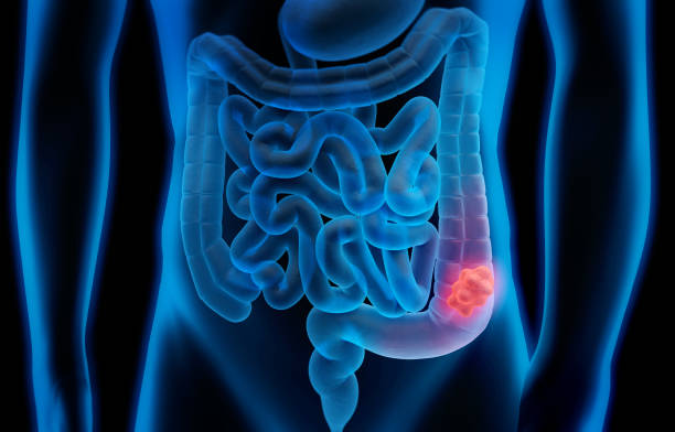
A condition known as colorectal cancer causes cells in either the colon or the rectum to grow out of control. Colon cancer is a short name for it at times. Colorectal cancer is the most prevalent cancer in Malaysian men (affecting about 15 in 100,000 people) and the second most common cancer in Malaysian women (affecting 11.1 in 100,000 people). It is also Malaysia’s third leading cause of cancer deaths. A polyp that develops cancer may eventually spread to the colon or rectum wall. There are numerous layers in the wall of the colon and rectum. Colorectal cancer develops across any or all of the other layers after beginning in the innermost layer (the mucosa).
Cancerous cells can spread through the wall and develop into lymph or blood arteries (tiny channels that carry away waste and fluid). They can then proceed to neighboring lymph nodes or to much further parts of the body. The depth to which a colorectal cancer penetrates the wall and if it has progressed outside the colon or rectum determine the stage (extent of dissemination) of the disease. Adenocarcinomas make up the majority of colon cancers. These tumors begin in the cells that produce the mucus that coats the rectum and colon. Almost always, when doctors discuss colorectal cancer, they usually refer to this kind.
The prognosis (outlook) for some subtypes of adenocarcinoma, like signet ring and mucinous, may be worse than for other subtypes of adenocarcinoma. Although there are many contributing factors, our cells’ deoxyribonucleic acid (DNA) is where the final modifications take place. DNA alterations or mutations can cause cells to proliferate out of control, and the expression or repression of particular genes can result in the growth of tumors.
Early-stage colorectal cancer is likely to be symptom-free and can be identified through active screening for colorectal cancer. Some signs and symptoms of colorectal cancer are:
- Blood in the stool
- Change in bowel pattern (diarrhoea or constipation)
- Unintentional weight loss
- Abdominal pain
- Vomiting
- Feeling that your bowel does not empty thoroughly
- Feeling fatigue
- Frequently having gas pains or cramps, feeling full or bloated
Colorectal cancer can be diagnosed through Colonoscopy, Biopsy, Imaging tests such as x-rays, computed tomography (CT) scans, magnetic resonance imaging (MRI) scan and positron emission tomography (PET) scan. Blood tests with complete blood count, tumour markers and liver enzymes. Treatment options for colorectal cancer are :
- Surgery – most colorectal cancers will have to be removed with surgery:
- Early stage: Laparoscopic approach (key-hole surgery) or polypectomy (removing polyps during a colonoscopy)
- More advanced stage: Open surgery to remove the cancer and surrounding colon tissues and lymph nodes (glands)
- Chemotherapy
- Depending on the stage of the cancer, the patient may require pre-operative or post-operative chemo-radiotherapy
- Surgery, combined with appropriate chemoradiotherapy, offers more than 90% cure rate in stage one disease. Advance and recurrent colorectal cancer patients may be treated with heated chemotherapy during the operation, a procedure called Hyperthermic Intraperitoneal Chemotherapy
- Radiation therapy (high-energy x-rays), Immunotherapy and Targeted drug therapy.
Precancerous polyps can be identified by screening tests and removed before they turn into cancer. Additionally, colorectal cancer screening tests might find the disease early, when treatment is most successful. Additionally, it is advised that screening for colon cancer should start around the age of 45 for people with an average risk of developing the disease. However, those who are more at risk, such as those with a history of colon cancer in the family, should think about screeninIt is the fourth most commonly diagnosed cancer and the second leading cause of cancer death.g early.

Lung cancer
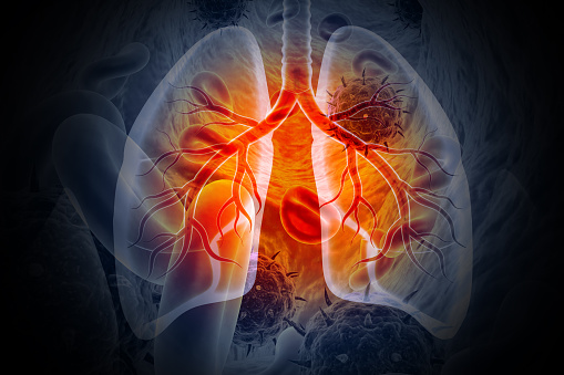
A type of cancer that appears in the lungs is called lung cancer. When normal cell division and growth processes go wrong, it results in the cells enlarging into a mass or tumor. Smoking is still the biggest factor contributing to lung cancer, even if some cases have no recognized cause. There are 2 main types of lung cancer and they are treated very differently.
Non-small cell lung cancer
Non-small cell lung cancer makes for about 80% to 85% of lung cancer cases. Large cell carcinoma, squamous cell carcinoma, and adenocarcinoma are the three primary subtypes of NSCLC. Because their prognosis outlooks and treatments are frequently similar, these subtypes, which originate from several types of lung cells, are categorized as NSCLC.
Adenocarcinoma: Adenocarcinomas start in the cells that would normally secrete substances such as mucus.This type of lung cancer occurs mainly in people who currently smoke or formerly smoked, but it is also the most common type of lung cancer seen in people who don’t smoke. It is more common in women than in men, and it is more likely to occur in younger people than other types of lung cancer. Adenocarcinoma is usually found in the outer parts of the lung and is more likely to be found before it has spread. People with a type of adenocarcinoma called adenocarcinoma in situ (previously called bronchioloalveolar carcinoma) tend to have a better outlook than those with other types of lung cancer.
Squamous cell carcinoma: Squamous cell carcinomas start in squamous cells, which are flat cells that line the inside of the airways in the lungs. They are often linked to a history of smoking and tend to be found in the central part of the lungs, near a main airway (bronchus).
Large cell (undifferentiated) carcinoma: Large cell carcinoma can appear in any part of the lung. It tends to grow and spread quickly, which can make it harder to treat. A subtype of large cell carcinoma, known as large cell neuroendocrine carcinoma, is a fast-growing cancer that is very similar to small cell lung cancer.
Small cell lung cancer
Compared to NSCLC, Small cell lung cancer tends to grow and spread more quickly. When diagnosed with SCLC, almost 70% of patients already had the disease disseminated throughout their bodies. This cancer tends to respond effectively to chemotherapy and radiation treatment because of how quickly it spreads. Unfortunately, the cancer will eventually come back for the majority of people.
Lung cancer does not initially show any observable signs or symptoms. When it has proceeded to a more severe level, symptoms become more noticeable. Here are a few possible lung cancer symptoms:
- Persistent cough, Coughing up blood
- Shortness of breath, Chest pain
- Hoarseness of voice, Unexplained weight loss over a short duration
- Bone pain, Headache
- Weakness or tired all the time, New onset of wheezing
- Chest pain that worsens with coughing or laughing
Lung cancer can be detected using a variety of different tests. In order to identify the origin and stage of your cancer and provide you with the most appropriate treatment, your doctor may carry out one or more of the following tests such as Chest x-ray , Computed tomography (CT) scan, Magnetic resonance imaging (MRI) scan, Positron emission tomography-CT (PET-CT) scan, Bronchoscopy to view the airways in the lungs and biopsy/cytology, Endoscopic ultrasound to see if cancer has spread to the lymph nodes located at the centre of the chest, Percutaneous lung biopsy using a long needle to take a lung tissue sample, Surgical biopsy to take a lung tissue sample, Mediastinoscopy to check the centre of the chest and if cancer has spread to the windpipe, Gene mutation testing and Bone scan to check if cancer from the lung has spread to the bone.
Treatment for lung cancer depends on its type and how much it has spread. Here are some ways in which lung cancer is treated:
- Chemotherapy: Special medication is circulated around the body to kill the cancer cells. Chemotherapy drugs can be taken orally or intravenously.
- Radiation therapy: High energy beams are aimed at the tumour or specific area to kill the cancer.
- Surgery: Your doctor performs an operation to remove the tumour and cancerous tissues.
- Targeted therapy: Drugs are used to stop cancer from growing and spreading.
Sometimes, a combination of the methods above is used. For example, your doctor might recommend chemotherapy to shrink a tumour before it is surgically removed. Early detection of lung cancer increases the chances of successful treatment. When a person smokes or has smoked in the past but shows no symptoms, it is advised that they have a lung cancer screening.

Nasopharyngeal cancer
Nasopharyngeal cancer, also referred to as nose cancer, is an uncommon kind of cancer that develops in the head and neck. Usually, it begins in the nasopharynx, or upper part of the throat. The base of the skull is where the nasopharynx is placed, near the roof of your mouth. When we breathe, the air moves from the nasal cavity through the throat and lungs. Nasopharyngeal cancer, also referred to as NPC, is a disease that is unique to Southeast Asia and has an incidence rate of up to 50 cases per 100,000 persons. Compared to the United States and other regions of the world, where there is a recorded incidence of fewer than 1 case per 100,000 persons, this is far lower.
Nasopharyngeal carcinoma (NPC)
Nasopharyngeal carcinoma is the most frequent type of nasopharynx cancer (NPC). Epithelial cells, which cover the surfaces of the body’s organs, are where carcinomas, a type of cancer, first appear.
There are different types of NPC. They all start from epithelial cells that line the nasopharynx, but the cells of each type look different when looked at closely in the lab:
- Keratinizing squamous cell carcinoma is the most common type in places with low rates of NPC, like the US.
- Non-keratinizing differentiated carcinoma is less common in areas with high rates of NPC and is often associated with the Epstein-Barr Virus (EBV).
- Non-keratinizing undifferentiated carcinoma is the most common type in areas with high rates of NPC and is often associated with EBV.
- Basaloid squamous cell carcinoma is rare and very aggressive.
The symptoms of nasopharyngeal cancer are similar to those of other, less serious medical illnesses, making it difficult to identify it in its early stages. Furthermore, in some cases cancer does not show many obvious symptoms until it is already advanced. Some notable symptoms of nasopharyngeal cancer include:
- A lump in the neck (most common symptom), Headache
- Double vision, Nose bleeds, Stuffy nose
- Unintentional weight loss, Swallowing problems
- Numbness at the lower region of the face, Hearing loss (usually happens in one ear)
- Trouble speaking or breathing, Facial pain, Ear infections, Ringing in the ears (tinnitus)
Nasopharyngeal cancer can be diagnosed through:
- Physical examination: The oncologist will ask detailed questions on your past medical history and closely examine your mouth, neck, throat, lymph nodes and head.
- Nasal endoscopy: A thin, flexible, and lighted tube known as an endoscope will be inserted through your nose and throat to look for any abnormalities.
- Biopsy: A sample tissue from the affected area is removed and examined under a microscope to find the presence of a tumour.
- Imaging tests:
- Positron emission tomography (PET) scan, Magnetic resonance imaging (MRI) scan, Computed tomography (CT) scan and Chest x-ray.
There is a specific treatment method suitable for every stage of Nasopharyngeal cancer. Some of these include:
- Radiation therapy: As nasopharyngeal cancer cells are sensitive to radiation, high-energy x-ray beams are used to destroy the cancer cells.
- Chemotherapy: Useful for cancers that have spread to different body parts. Anti-cancer drugs are given intravenously or orally.
- Targeted drug therapy: This is often combined with radiation therapy and chemotherapy. Nasopharyngeal cancer patients may benefit from cetuximab injections (synthetic version of an immune system protein).
- Surgery: Nasopharynx is a tricky area to operate, however in some in certain cases the tumour can be surgically removed. Surgery can also be used to remove lymph nodes that are not responding to other treatments.
Early identification can lead to improved patient outcomes. Doctors may offer screening for people who are considered at high risk for nasopharyngeal cancer.

Liver cancer
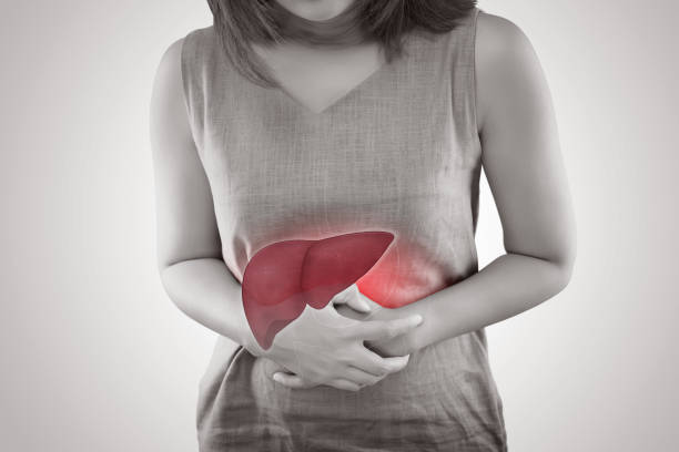
One kind of cancer that begins in the liver is known as liver cancer. Primary liver cancer is a term used to describe cancer that begins in the liver. There are various primary liver cancer types.
Hepatocellular carcinoma (HCC)
Hepatocellular cancers can have different growth patterns such as some develop as a single tumour that spreads. The disease does not initially spread to other areas of the liver. Instead of simply one large tumour, a second form appears to begin as numerous little cancer nodules spread throughout the liver. People with cirrhosis are the ones who experience this the most frequently (chronic liver damage)
Intrahepatic cholangiocarcinoma (bile duct cancer)
Cholangiocarcinoma within the liver is responsible for 10% to 20% of all cancers. These tumours begin in the liver’s tiny bile ducts, which are tubes that deliver bile to the gallbladder. But the majority of cholangiocarcinomas really begin in bile ducts that are external to the liver. Cholangiocarcinomas are often treated in the same way, despite the fact that the remainder of this information focuses mostly on hepatocellular malignancies. See Bile Duct Cancer for more information on this particular type of cancer.
Angiosarcoma and hemangiosarcoma
These are rare cancers that start in the cells lining the liver’s blood veins. These cancers are more likely to develop in those who have been exposed to vinyl chloride or thorium dioxide (Thorotrast). Some additional cases are thought to be caused by genetic hemochromatosis, exposure to arsenic or radium, or both. It is impossible to pinpoint a likely source in around half of all instances. By the time these tumours are discovered, they have typically gone too far to be surgically removed. Although these cancers are typically exceedingly difficult to treat, chemotherapy and radiation therapy may help slow the disease. These cancers receive similar treatment as other sarcomas.
Hepatoblast
This extremely rare type of cancer typically affects children under the age of four. Hepatoblastoma cells resemble fetal liver cells in appearance. tumoursThe tumors are difficult to treat if they have spread outside the liver, but about 2 out of 3 children with these tumours are successfully treated with surgery and chemotherapy.
Secondary liver cancer (metastatic liver cancer)
When cancer is discovered in the liver, it usually did not start there but rather had spread (metastasized) from the pancreas, colon, stomach, breast, or lung. This cancer is referred to as a secondary liver cancer since it has spread from its primary (original) site. Based on the primary site, these cancers are given names and treatments (where they started). For instance, lung cancer that has gone to the liver is referred to as lung cancer has spread to the liver rather than liver cancer. It is also treated as lung cancer.
Benign liver tumors
Although benign tumors can occasionally become large enough to be problematic, they do not invade neighboring tissues or spread to other parts of the body. Surgery is typically able to cure the patient if they need to be treated.
Hemangioma
Hemangiomas, the most common form of benign liver tumour, begin in blood vessels. The majority of liver hemangiomas are asymptomatic and do not require medical attention. However, some of them could bleed and require surgery to be removed.
Hepatic adenoma
A benign tumor that originates from hepatocytes is called a hepatic adenoma (the main type of liver cell). The majority are symptomless and do not require treatment. However, some eventually result in symptoms like pain, a bulge in the abdomen (the area of the stomach), or blood loss. The majority of specialists will typically suggest surgery to remove the tumor if possible due to the risk that it could rupture (resulting in significant blood loss) and the minor possibility that it could ultimately develop into liver cancer. Taking some medications may make these tumors more likely to develop. Birth control pill users have a greater risk of developing one of these tumors, although it is exceptionally rare. These tumors can also occur in men who use anabolic steroids. When these medications are withdrawn, adenomas may get smaller.
Focal nodular hyperplasia
Several different cell types make up the tumor-like growth known as focal nodular hyperplasia (FNH) (hepatocytes, bile duct cells, and connective tissue cells). Despite being benign, FNH tumors can nevertheless present with symptoms. They can be challenging to distinguish from actual liver malignancies, so when the diagnosis is murky, doctors occasionally remove them. Hepatic adenomas and FNH tumors affect women more frequently than they do men.
In the early stages of liver cancer, symptoms frequently do not show up. But as the illness worsens, more signs and symptoms will become obvious. Some of the symptoms are :
- Unusual weight loss, Discomfort in the right upper side of the abdomen
- A hard lump in the right abdomen just below the rib cage, Nausea and vomiting
- Loss of appetite, Jaundice (yellowing of the skin and white part of the eyes)
- A swollen abdomen, Fatigue, Dark-coloured urine
Upon experiencing some of the symptoms mentioned above, one should immediately consult an oncologist. As they might recommend some of the following tests to rule out cancer such as :
- Physical examination: This is one of the preliminary tests done by the physician to check for liver cancer signs and symptoms.
- Biopsy: A small amount of tissue from the affected region is removed and observed under a microscope to check for abnormal cells.
- Blood tests: High alpha-fetoprotein (AFP) levels in the blood may suggest liver cancer.
- Ultrasound scan: Sound waves are used to look for the tumours.
- Computed tomography (CT) scan: This specialized X-ray captures comprehensive images of your liver, providing information on the size and location of liver tumours.
- Magnetic resonance Imaging (MRI): An MRI uses magnetic fields to produce detailed images of the internal parts of the body.
- Laparoscopy: In this procedure, a lighted, thin, flexible tube known as the laparoscope is inserted into the abdominal region through a small incision to check the liver and its surrounding tissues.
Here are some ways in which liver cancer is treated:
- Cryotherapy: Freezing the tumour/cancer cells
- Chemotherapy: Drugs are administered to kill the cancer cells
- Radiofrequency ablation: The use of electric current to destroy the tumour
- Radiation therapy: Specific radiations are used to kill the cancer cells
- Immunotherapy: The body’s immune system is stimulated to destroy the cancer cells
- Targeted drug therapy: The drug targets a specific abnormality in the cancer cells to destroy them
- Liver transplant surgery: Replacing the diseased liver with a healthy one from a donor
- Partial hepatectomy: Removing the liver cancer and a small part of the liver tissue
For those with a higher risk of getting the disease, such as those with hepatitis B or C infection and liver cirrhosis, screening for liver cancer is advised. Every six months, a blood test and an abdominal ultrasound are part of the liver cancer screening process.

Conclusion
Cancer is frequently linked to death, pain, suffering, and incurability. A cancer diagnosis is frequently accompanied by a greater fear of dying than many other illnesses. A person may wonder how bad their disease is and whether they will survive it after receiving a cancer diagnosis. Prognosis is an assessment of how a condition will impact you.
There are many factors that affect your prognosis, such as:
- Type of cancer and location in your body
- Cancer stage (size of the tumour and if it has spread to other parts of your body)
- Cancer grade (how abnormal the cells are)
- Certain characteristics of the cancer cells
- Your age
- Your health status
- Your response to cancer treatment
One of the main determinants of cancer survival is the stage of cancer at diagnosis. Survival by cancer type and stage of diagnosis by cancer type and stage of diagnosis
|
Cancer |
Early stage (Stage 1) |
Late stage (Stage 4) |
|
Female breast |
87.5% |
23.3% |
|
Colorectal |
75.8% |
17.3% |
|
Lung, trachea and bronchus |
37.1% |
6.3% |
|
Nasopharyngeal |
63.7% |
26.9% |
|
Liver |
20.4% |
9.2% |
Infographic from : Gleneagles hospital


